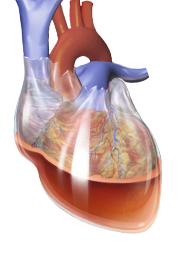
Cardiac Tamponade
The Pericardium is made up of 2 layers; the visceral and parietal pericardium. These layers contain a small amount of fluid (< 50 mls) between them for lubrication. This lubrication allows the heart to beat w/o generating much friction. In a pericardial effusion, this potential space between the layers becomes more filled, therefore putting pressure on the heart and making it more difficult to expand during diastolic filling.
Pericardial effusion - Excess fluid collects in pericardial sac. This fluid can be:
Transudative - low protein
Exudative - high protein, and associated w/inflammation
Blood
Pus
Gas - associated w/bacterial infections
Cardiac Tamponade - Effusion becomes large enough to increase the intra-pericardial pressure, therefore squeezing the heart and reducing it’s effectiveness during diastole AND systole → Reduced CO. This requires rapid drainage with Pericardiocentesis.
Causes
Transudative effusion - Due to an increased venous pressure, which reduces drainage from the pericardial cavity. This is typically caused by Congestive HF and Pulmonary HTN.
Exudative effusion - Due to inflammation of the pericardium (pericarditis). This can be caused by:
Infection
Autoimmune e.g. SLE, RA
Injury to pericardium e.g. post-MI, surgery, trauma
Uraemia secondary to renal impairment
Cancer
Medications e.g. methotrexate
Haemopericardium - Due to rupture of the heart/aortic root. This can be caused by Trauma, MI, Type A Aortic Dissection.
Presentation
A rapid collection of fluid can quickly cause haemodynamic instability and collapse (Cardiac tamponade), whereas a slower collection of fluid may initially be asymptomatic and, as pressure rises, the symptoms develop.
Pericardial effusion will present with:
Chest pain
SOB
Orthopnoea
Feeling of fullness in chest
Compression of surrounding structures, leading to:
Phrenic nerve → Hiccups
Oesophagus → Dysphagia
Recurrent laryngeal nerve → Hoarse voice
Cardiac Tamponade presents with Beck’s Triad – Hypotension, Muffled heart sounds, Raised JVP.

Signs O/E include:
Muffled heart sounds
Pulsus Paradoxus (large fall in BP on inspiration)
Hypotension
Raised JVP
Fever and pericardial rub with pericarditis
Investigations
Echo
Pericardiocentesis - Fluid analysis on the pericardial fluid to determine the underlying cause - Protein (distinguishes between transudative and exudative)
ECG – Small QRS complexes
CXR – Large globular heart
Management
If the patient is haemodynamically unstable, they need an urgent Pericardiocentesis. Complications that occur with this procedure are pneumothorax, myocardial damage etc. – CXR should be done post-procedure to exclude this.
Important Links: