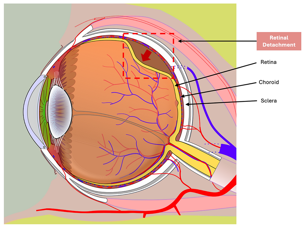
Retinal Detachment
Retinal detachment is where the retina separates from the vascularised choroid layer, usually due to a tear, therefore allowing the vitreous fluid to escape behind and fill the space between the layers.
Risk factors:
Trauma
Posterior vitreous detachment
Diabetic retinopathy
Older age
FHx
N.B. A viterious detachment won't present with visual loss, but retinal detachment will.
Presentation
Sudden painless and progressive vision loss - Usually starts peripherally and progresses towards the centre as the tears extends i.e. like a curtain
Floaters
Flashes
Blurred vision

Management
Managing the retinal tear - Laser therapy or Cryotherapy, which creates adhesions between the retina and choroid
Managing the retinal detachment:
Vitrectomy – Vitreous attached to the retinal tear is removed, and a gas or oil bubble is added to span and close the tear until a scar forms
Scleral buckling - Silicone ‘buckle’ is placed on the scleral surface. This indents the outer eye to make contact with the detached retina.
Pneumatic retinopexy – Small, expansile gas bubble is injected into the vitreous in the right area to flatten the detached retina against the choroid

Important Links:
https://www.nhs.uk/conditions/detached-retina-retinal-detachment/
https://cks.nice.org.uk/topics/retinal-detachment/
https://bestpractice.bmj.com/topics/en-gb/651 “Diagram of a human eye (horizontal section section of the right eye)” © Jmarchn CC BY-SA 3.0 (https://creativecommons.org/licenses/by-sa/3.0/)
“Snapshot of 3-port 23gauge vitrectomy procedure” © Drmandar CC BY-SA 3.0 (https://creativecommons.org/licenses/by-sa/3.0/)
“Cross section of a human eye, showing a scleral buckle (blue), which brings the choroid (yellow) into contact with a detached retina (red).” © Erin Silversmith, Delta G, RexxS CC BY-SA 3.0 (https://creativecommons.org/licenses/by-sa/3.0/)