Lower Limb Disorders
Hip Fracture
This presents with:
Shortened, abducted, and externally rotated leg
Pain
Inability to weight bear
Risk factors - Increasing age, Osteoporosis
It's classified into whether the fracture is Intracapsular and Extracapsular. Intracapsular requires more urgent surgery as it carries a higher risk of avascular necrosis.

Total hip replacement is a common management option, however a complication of this is Posterior hip dislocation. Here, patients will have a shortened, but internally rotated leg.
N.B. For hip -> External rotation = Fracture. Internal rotation = Dislocation.
Meniscal Tears
This often occurs during twisting movements. It presents with:
Pop sound during initial injury
Pain, Swelling, Stiffness
Reduced range of motion
Locking of knee
Knee instability
Investigations:
McMurray’s and Apley Grind tests
MRI
Arthroscopy

ACL Injury
The ACL functions to stop the tibia from sliding forwards. An injury of this often occurs during sudden deceleration or twisting of the knee. It presents with:
Pop sound during initial injury
Pain, Swelling, Stiffness
Reduced range of motion
Knee instability
Investigations:
Anterior Drawer test
MRI
Arthroscopy

Achilles Tendinopathy
Inflammation of the achilles at the mid-point or insertion point. It's caused by tearing/straining of the tendon fibres over a long period of time. It presents with pain, stiffness, tenderness, swelling, and thickening of the tendon.
Risk factors - Sport, Inflammatory conditions, DM
An Achilles tendon rupture always needs to be ruled out by doing a Simmonds’s calf squeeze test.

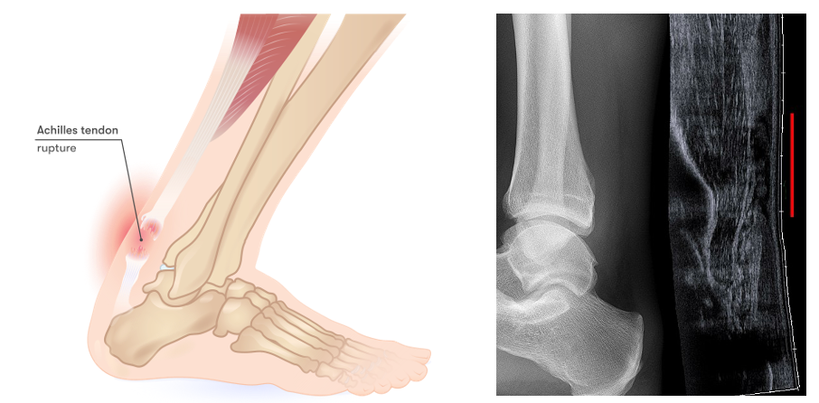
Achilles Tendon Rupture
This presents with:
Sudden onset pain
Snapping sound during initial injury
Feeling of something hitting them at the back of the leg
Weakness of plantarflexion
Investigations:
Simmond’s calf squeeze test
US
Plantar Fasciitis
This presents with a gradual onset of pain, which is worse with pressure e.g. walking or standing for long periods.

Ankle Fracture
This is sub-divided using the Weber Classification. This helps us know how unstable the fracture is and guides how we manage it:
Type A - Below syndesmosis
Type B - At level of syndesmosis
Type C - Above syndesmosis
Management:
Stable - Type A - Managed with Plaster of Paris (POP) cast
Unstable - Type B and C - Managed with Surgery

Important Links:
https://orthoinfo.aaos.org/en/diseases--conditions/hip-fractures/
https://orthoinfo.aaos.org/en/diseases--conditions/meniscus-tears/
https://orthoinfo.aaos.org/en/diseases--conditions/anterior-cruciate-ligament-acl-injuries/
https://orthoinfo.aaos.org/en/diseases--conditions/achilles-tendinitis/
https://orthoinfo.aaos.org/en/diseases--conditions/achilles-tendon-rupture-tear-video/
https://orthoinfo.aaos.org/en/diseases--conditions/plantar-fasciitis-and-bone-spurs
https://orthoinfo.aaos.org/en/diseases--conditions/ankle-fractures-broken-ankle/
“Medical illustration of achilles tendon rupture.” © InjuryMap CC BY 4.0 (https://creativecommons.org/licenses/by/4.0/)
“Achilles tendon rupture seen at sonography: discontinuity over several centimeters (red line). No fracture or avulsion (radiograph).” © Hellerhoff CC BY-SA 3.0 (https://creativecommons.org/licenses/by-sa/3.0/)